
If you have to bring your pet to your vet's for diagnostic imaging, you may have a lot of questions: How are X-rays, ultrasounds and CT scans used on dogs and cats? What is the process? Do they have to be sedated? Our Westport vets explain the ins and outs of routine diagnostic imaging for dogs and cats.
How does diagnostic imaging help my pet?
X-rays, ultrasounds and CT scans are frequently used in both human healthcare and veterinary healthcare to help diagnose health conditions.
Electromagnetic radiation and other technologies are used in routine diagnostic imaging procedures for dogs and cats. They capture extremely detailed images of your pet's bones, soft tissues and other internal structures so your vet can offer an accurate diagnosis and plan effective treatments.
In this post, we'll explain each of the diagnostic tools listed above and how they are used with pets.
X-Rays, Ultrasounds & CT Scans for Dogs & Cats
Wondering how this equipment is used on dogs and cats? Here are brief explanations for each:
Radiography (Digital X-Rays)
With a digital X-ray (radiograph)s for cats or dogs, we use very low doses of radiation to examine the inside of the body to reveal important information that might not be easily viewed from the outside.
This procedure is used to evaluate organs and bones, and to diagnose conditions such as spinal cord diseases, arthritis, broken bones, bladder stones and some tumors.
Because radiation levels are so low, even very young pets and pregnant females are able to undergo radiography.
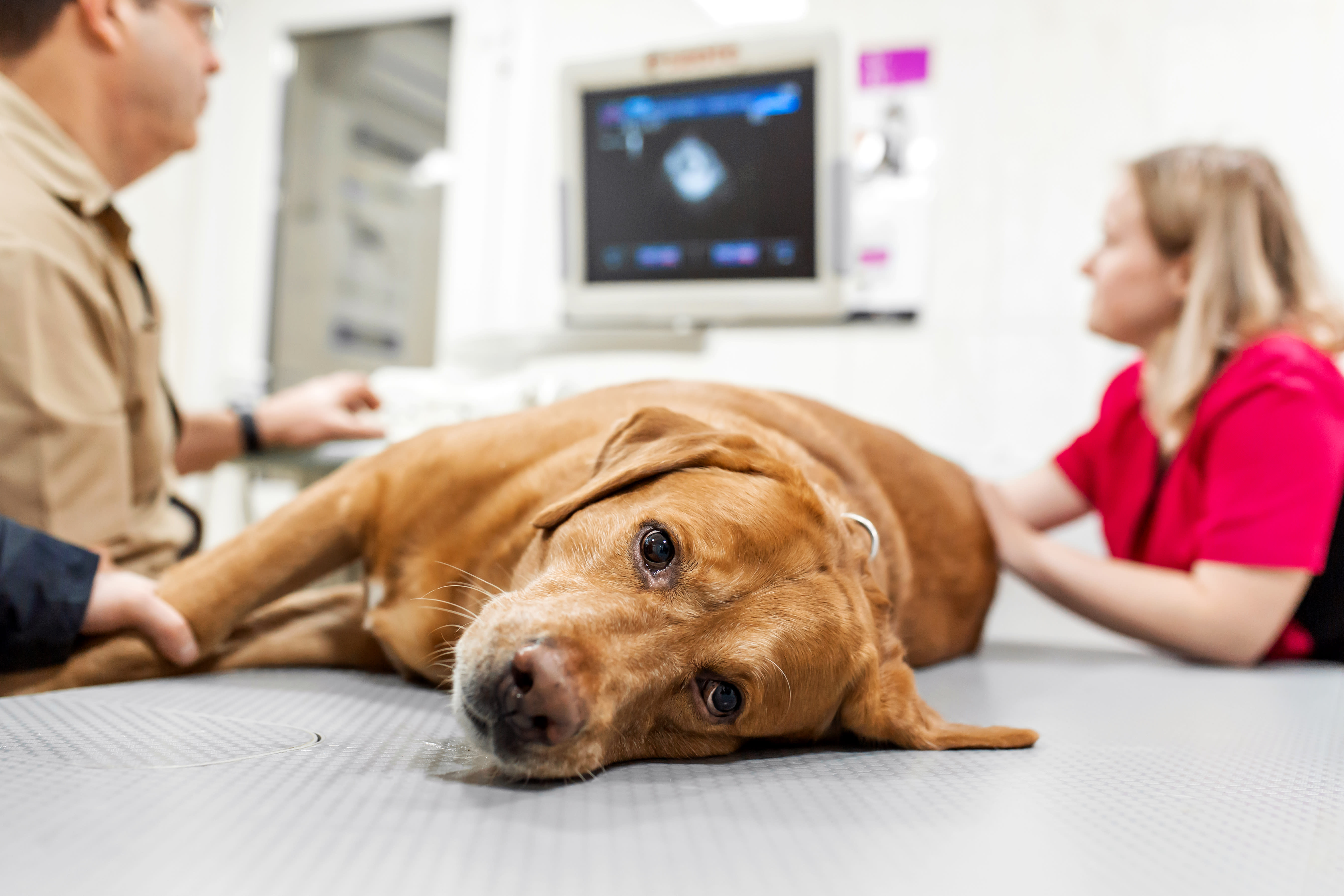
Ultrasound
This non-invasive diagnostic imaging technique uses sound waves that are transmitted into an animal's body, which then produce an image your vet can review to assess the internal organs and their architecture.
With ultrasound, soft tissue masses or foreign bodies can be distinguished from fluid - which can be difficult to do with a digital X-ray. If your pet has developed a tumor or ingests something he or she shouldn't have, ultrasound can help locate and characterize the objects. This technique is also useful for seeing blood in the abdomen and surrounding the heart.
CT Scans
Often referred to as a Cat Scan or CT scan, computed tomography is useful when assessing the nasal passage, sinuses, lungs, thorax, ears, abdomen and some orthopedic areas.
While CT scans are not offered here, we can refer you to a specialist for one if required.
Your vet might recommend a CT scan if your pet has any condition ranging from lung disease to pulmonary fibrosis, metastatic cancer (before surgery), tumors or masses in the chest cavity, disease in the nasal cavity, trauma to the spine or pelvis, vascular anomalies or orthopedic developmental disease (elbow dysplasia).
CT scans offer improved differentiation between bones and soft tissues than conventional X-rays (radiographs). The animal lies on a table, which slowly advances into the part of the machine performing the scan. CT Scans for dogs or cats only take a short amount of time.
An X-ray tube rotates around the patient to record X-rays from several angles (the suspected health issue will determine the number of images captured) to create slices. The slices are then stacked together to produce a 3D image of your pet without superimposition of other tissues or organs.
X-rays and CT scans are both non-invasive and not painful for either cats or dogs. The sound waves generated by ultrasounds are also non-invasive, and not harmful or painful.
Will my pet need to be sedated for diagnostic imaging?
For an X-ray, the answer to this question depends on whether your pet is calm, in pain, and able to lay in a comfortable position for the duration of the X-ray. Anesthesia may also be recommended if the X-ray needs to capture images of the skull, teeth or spine.
Since CT scans require a pet to be completely still, heavy sedation or general anesthesia is required. Your pet's vital signs will be closely monitored while the scan is performed.
If biopsies need to be done before an ultrasound, your pet will require a heavy sedative or short-acting anesthetic to help them relax while the vet performs the procedure and to avoid potential complications. Your vet will notify you if this is required.
How do I get my results?
Our vets can review results from digital X-rays and ultrasounds in-house and are often able to provide immediate diagnoses for health issue. In some cases, they may need to be sent to a veterinary radiologist for further consultation. If you are waiting for news about CT Scans for which you may be referred to a specialist, we will likely receive the results and review them with you before planning treatment.
Note: The advice provided in this post is intended for informational purposes and does not constitute medical advice regarding pets. For an accurate diagnosis of your pet's condition, please make an appointment with your vet.
